COREX Case Studies
Explore how surgeons are using COREX to harvest cancellous bone graft using a minimally invasive technique.
Discover How Easy it Is To Make Autologous Bone Your First Choice for Bone Graft
A minimally invasive technique combined with COREX addresses the traditional issues that contribute to harvest site morbidity including incision size, soft tissue stripping, and cortical disruption.
Case Study 1: Distal Tibia Harvest
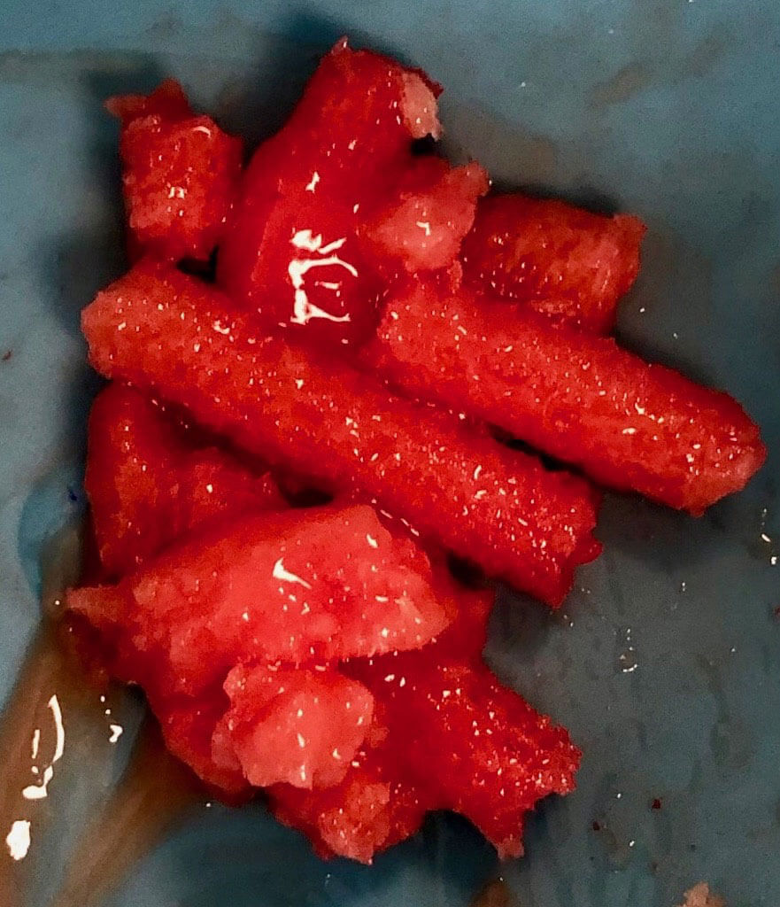
Figure 1. Intraoperative photograph of COREX minimally invasive bone harvester being utilized in the distal tibia (A) to harvest cores of autogenous bone graft (B).
Intraoperative image intensification images demonstrating creation of cortical access window (C) and the COREX device being passed in multiple angles (D, E, F) through the distal tibial metaphyseal bone for harvest.

Figure A
COREX-CS-1b
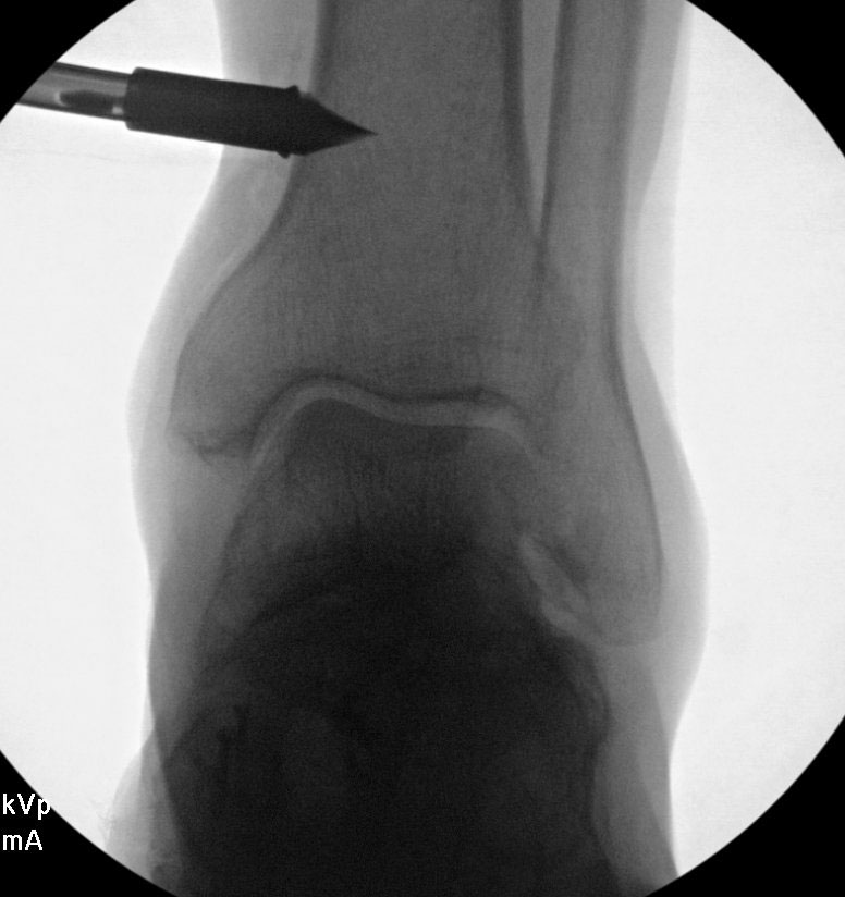
Figure C
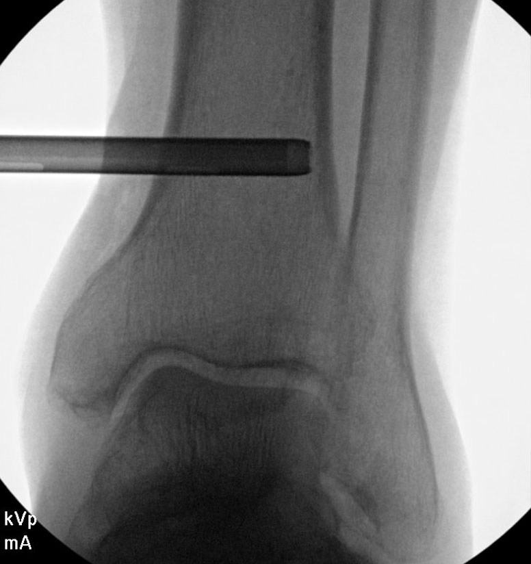
Figure D
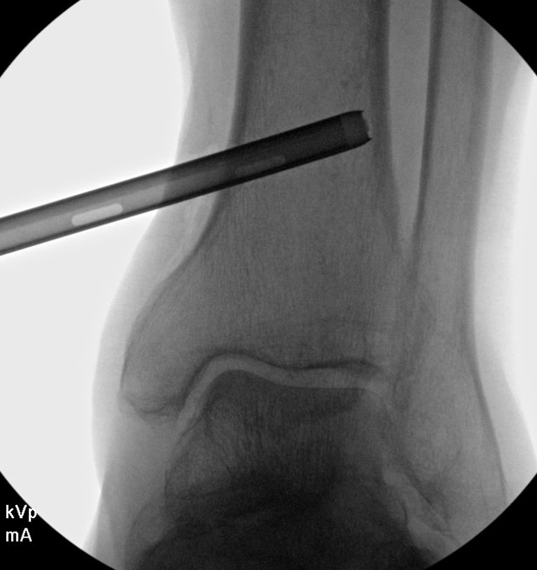
Figure E
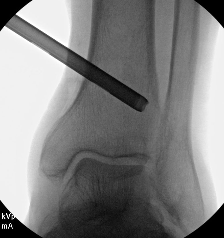
Figure F
Case Study 2: Calcaneus Harvest
Figure 2. Intraoperative image intensification demonstrating autogenous bone graft harvest site within the calcaneus (A) after using the COREX minimally invasive bone harvester to produce multiple cores of autogenous bone (B) to augment a great toe joint arthrodesis.
Intraoperative photograph of autogenous bone graft employed in the great toe joint arthrodesis site (C) with overlying fixation.
Postoperative weightbearing AP (D) and lateral (E) foot radiographs demonstrating complete healing of arthrodesis site and no complications at calcaneal bone graft harvest site.

Figure A
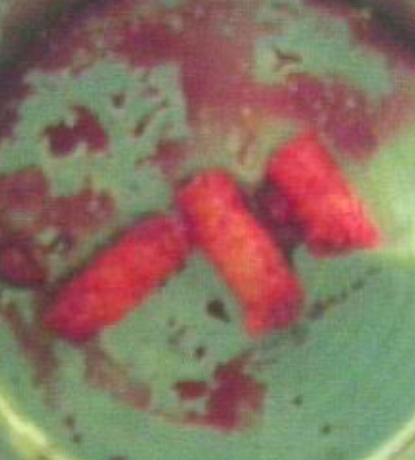
Figure B
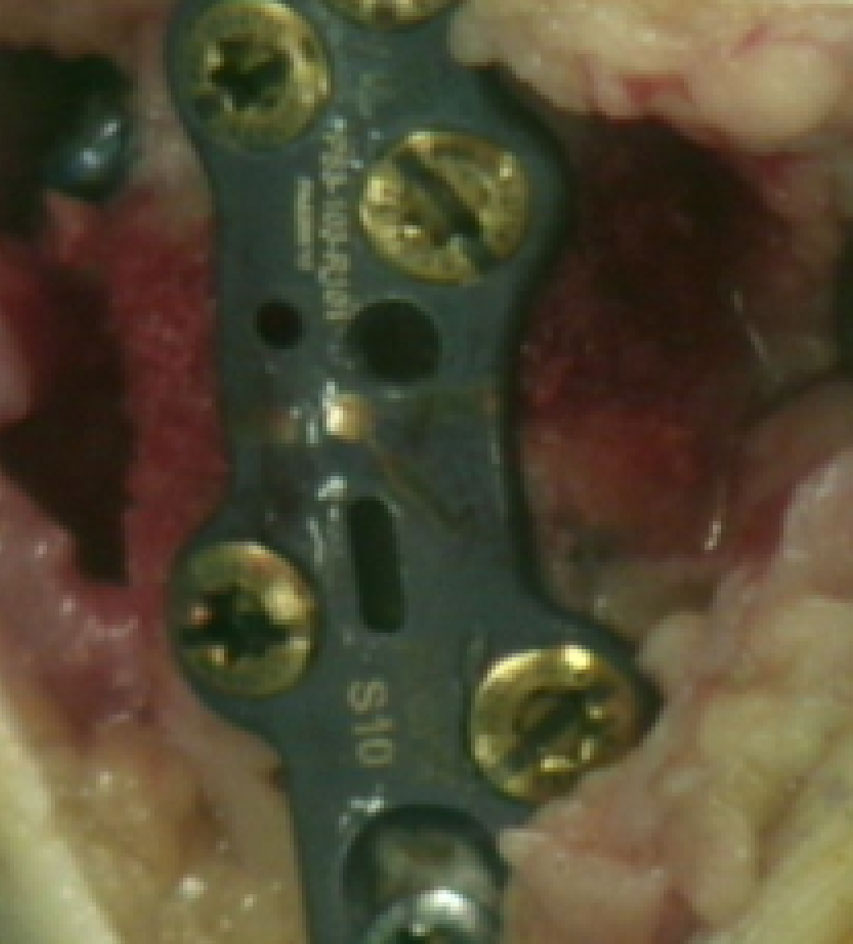
Figure C
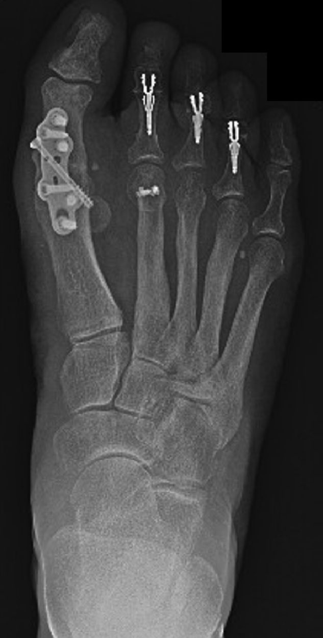
Figure D
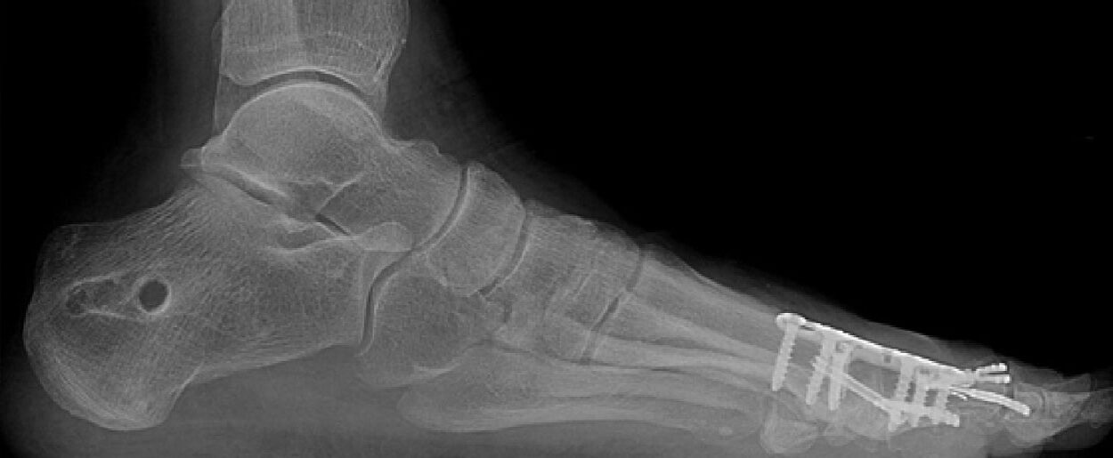
Figure E
Autologous Bone Cases and Publications

Autologous Bone Grafting in Trauma and Orthopaedic Surgery: An Evidence-Based Narrative Review
Download PDF

Percutaneous Harvesting of Cancellous Bone Graft: A Surgical Technique Harvesting from the Calcaneus Using COREX
View Video